 Vi
Vi 
A Danish patient suffered a haemorrhagic stroke, experiencing severe headaches and uncontrollable vomiting. Fortunately, he was quickly taken to FV Hospital, where the medical team promptly diagnosed his condition and performed a life-saving neurovascular intervention, avoiding the need for surgery.
A Life-Saving Journey
The doctors at FV Hospital’s Accident & Emergency Department swiftly conducted tests to assess the patient’s condition. An MRI scan revealed bleeding in the brainstem. Since the brain haemorrhage was not caused by trauma or impact, a follow-up CT scan confirmed a rare diagnosis: a dural arteriovenous fistula with a large 25mm venous aneurysm that had ruptured. Immediate intervention was critical, as the patient’s life was at imminent risk.
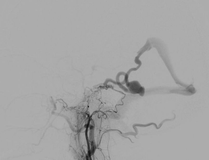
Without hesitation, the multidisciplinary medical team, including the Accident & Emergency, Anaesthesiology & ICU, and Imaging & Interventional Radiology departments, held an urgent consultation. Dr Huynh Huu Danh, Specialist Level I and expert in Imaging and Interventional Radiology at FV Hospital, assessed the case as a rare dural arteriovenous fistula with a large ruptured venous aneurysm, posing a risk of continued bleeding and a serious threat to the patient’s life.
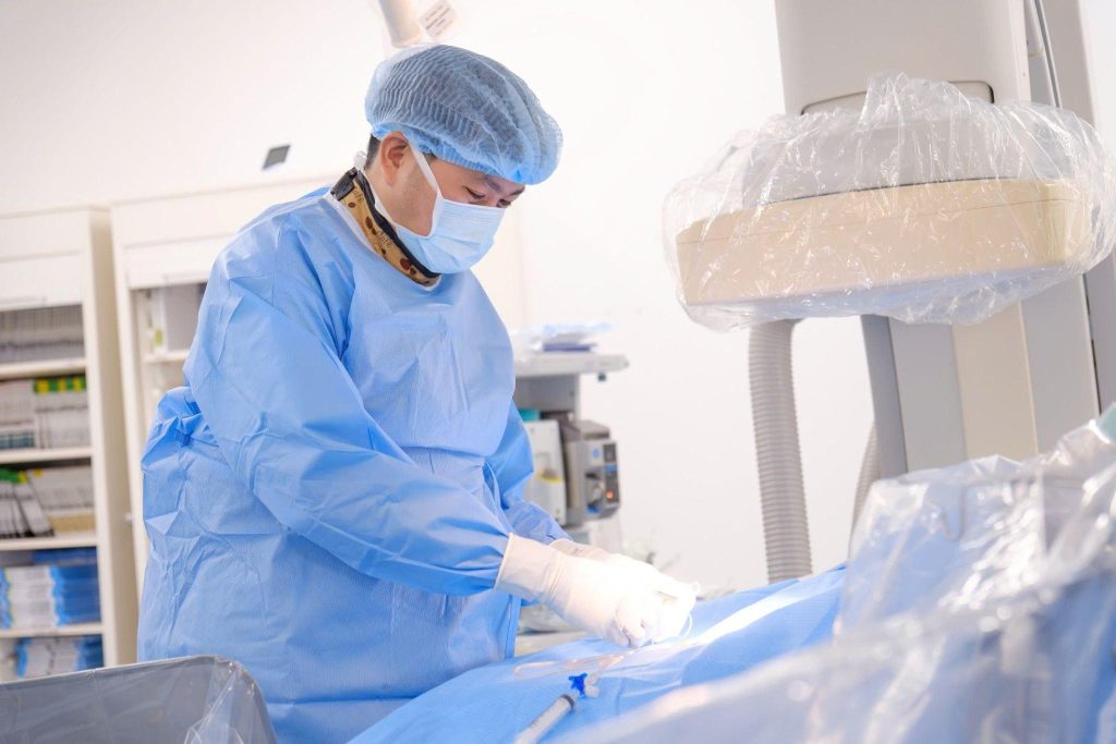
Instead of traditional open craniotomy surgery, the doctors opted for an endovascular intervention procedure. This is an advanced, minimally invasive treatment method known for its high effectiveness but requiring significant precision and expertise. The patient was immediately transferred to the Cathlab, where Dr Huynh Huu Danh personally performed the procedure with the support from the Digital Subtraction Angiography (DSA) system.
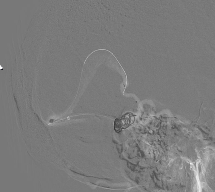
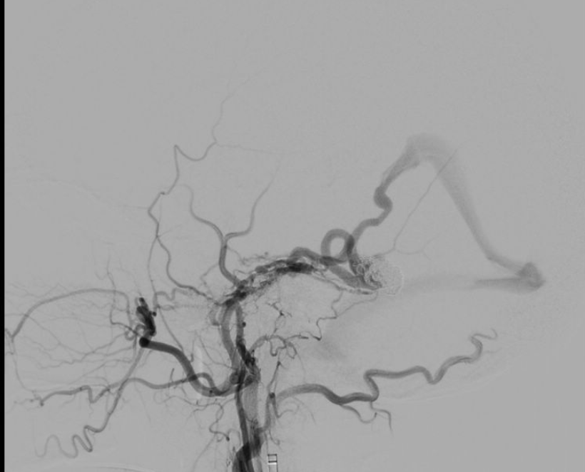
Dr Danh explained that due to the aneurysm’s location being difficult to access, he had to navigate the catheter through a vein instead of an artery. “If intervening through an artery is like driving on a smooth, paved road, then intervening through the vein is like crossing rugged, winding terrain, which carries a higher risk of complications,” Dr Danh compared.
Through a small incision in the thigh, Dr Danh carefully guided a catheter and micro-guidewire through the vein to reach the aneurysm in the brain. A total of 12 coils (metallic spiral wires) were placed inside the aneurysm to “patch” the damaged area and block the blood flow through the rupture.
After just a few hours, the patient stabilized, with no further nausea and a reduced headache. MRI scans in the following days confirmed that the brain haemorrhage had not progressed and was gradually resolving. Mr F. was discharged after two weeks of monitoring.
A Very Rare Condition, But a Silent Killer
Dural arteriovenous fistula is a very rare condition that can be caused by infection, trauma, or may be congenital. When accompanied by a venous aneurysm, it becomes even rarer, with a risk of brain haemorrhage. Patients often show no symptoms until the fistula or aneurysm ruptures, leading to a haemorrhagic stroke that can be life-threatening.
To detect cerebrovascular diseases that may lead to stroke at an early stage, the most effective method is an MRI brain scan. At FV Hospital, a stroke screening programme is available, where patients are directly examined by a neurointerventionalist and cardiologist. The screening includes evaluation of organ function, magnetic resonance angiography (MRA), and carotid artery imaging.
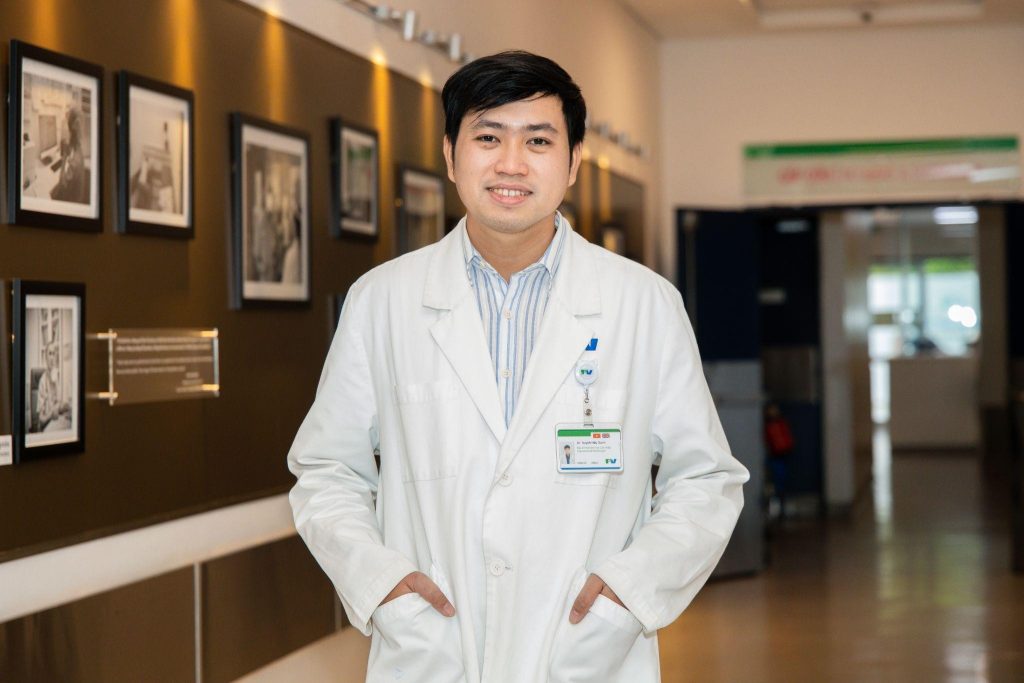
Treating dural arteriovenous fistulas or brain aneurysms with endovascular intervention offers the advantage of being minimally invasive while achieving a high success rate. However, this technique must be carried out in a modern Cathlab and requires specialized doctors in both endovascular and neurointerventional procedures. FV Hospital is equipped with a modern Cathlab and a DSA imaging system, which delivers high-resolution vascular imaging to enhance precise procedures and reduce intervention time. Dr Huynh Huu Danh, Specialist Level I, is one of the experienced endovascular specialists, qualified to perform these procedures on both adults and children.
| FV Hospital officially applies Health Insurance for Neurointerventional Radiology procedures with support of up to 50 million VND. Covered procedures include chemical embolization for liver cancer (TACE), uterine fibroid embolization, prostate embolization, embolization for various types of tumours (liver, kidney, bone, soft tissue, vascular tumours, vascular malformations), carotid artery thrombectomy, brain aneurysm treatment, brain vascular malformation intervention, and embolization for dural arteriovenous fistulas. For more information, please contact 028 5411 3333 or visit us directly at 06 Nguyen Luong Bang, District 7, Ho Chi Minh City. |