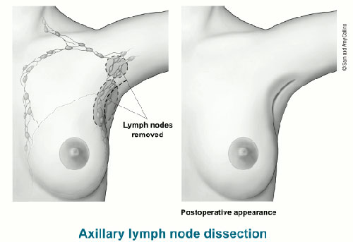
After total lymph node removal surgery, breast cancer patients may suffer from severe complications, such as arm puffiness and numbness. Patients at Hy Vong Cancer Care Centre that undergo sentinel lymph node biopsy procedure do not have to worry about experiencing complications in their arms.
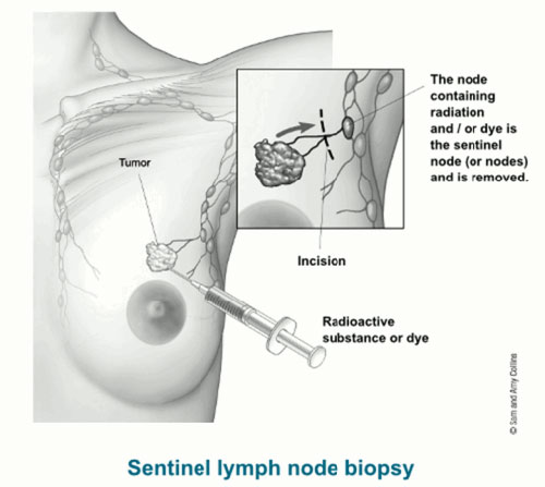
Sentinel lymph nodes acts as guards. In breast cancer patients, sentinel lymph nodes are the first lymph nodes that cancer cell encounter when breaking away from the tumour. Cancer cells are typically found in sentinel lymph nodes before spreading to other lymph nodes. Therefore, in modern breast cancer treatment, physicians will look for sentinel lymph nodes and perform a local biopsy. If sentinel lymph nodes are found “clear”, the possibility that other lymph nodes contain cancer cells is nearly zero. Patients then only need to undergo surgery to remove the tumour without dissection of all axillary lymph nodes as in traditional surgical treatment procedure.

However, a sentinel lymph node biopsy is recommended for patients who are diagnosed with early breast cancer and have relatively small tumours. Clinical studies show that after almost five years, patients who have just the sentinel nodes removed have equal survival and non-recurrence rates as patients who have total axillary lymph nodes removal.
Hy Vong Cancer Care Centre at FV Hospital is the first clinical facility in Vietnam putting radio-isotope angiography machines to locate sentinel lymph nodes into use; the team has been using this methodology to detect early breast cancer metastasis since May 2010.

As the sentinel lymph nodes are located, a local biopsy is conducted in an operating theatre to identify whether metastasis has occurred. A negative biopsy result means that the sentinel lymph nodes contain no cancer cells. In this case, tumour removal without axillary lymph nodes dissection is recommended to patients. If the biopsy result is positive, a tissue specimen is sent to Laboratory for histological analysis to verify the test result before the next step in the patient’s treatment plan is developed.
The utilisation of radio-isotope angiography is an effective means of support for physicians during cancer care and ensures that an accurate diagnosis and optimal treatment plan for each individual patient can be reached; this method also satisfies the quality care requirements and standard treatment protocols widely applied in occidental countries.

 Vi
Vi 


