WHAT IS ULTRASOUND?
Diagnostic ultrasound, also called sonography or diagnostic medical sonography, is an imaging method that uses high-frequency sound waves to produce images of structures within your body. The sound waves are recorded and displayed as a real-time visual image. No ionising radiation (X-ray) is involved in ultrasound scanning.
In most ultrasound examinations, a transducer – a lightweight device which produces sound waves – is placed on the patient’s skin. There are also special transducers which can be put into the vagina or rectum to image these areas of the body.

WHAT ARE THE COMMON USES OF ULTRASOUND?
Ultrasound is used:
- For imaging abdominal organs (liver, gallbladder, spleen, pancreas, kidneys, bladder), pelvic organs (prostate, testicles, uterus and ovaries) and superficial organs (breast, thyroid, joints of shoulder or ankle);
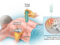
- To guide interventional procedures such as needle biopsies, Fine Needle Aspiration (FNA), in which a needle is used to sample cells from an organ for laboratory testing;
- To view the uterus and ovaries of a pregnant woman and assess her foetus.
Doppler ultrasound is a special technique used to examine flow in blood vessels. It allows the doctor to see and evaluate blockages, such as clots, and build-up of plaque inside the vessels.
WHAT ARE THE BENEFITS, RISKS AND LIMITATIONS?
Benefits
- Ultrasound scanning is non-invasive (no needles or injections in most cases) and is usually painless.
- Ultrasound is widely available and easy to use.
- Ultrasound imaging uses no ionising radiation and is the preferred image modality for diagnosis of pregnant women.
- Ultrasound provides real-time imaging, making it a good tool for guiding minimally invasive procedures such as needle biopsies.
- Ultrasound images can visualise structures, movement and live function in the body’s organs and blood vessels.
Risks
For standard diagnostic ultrasound there are no known harmful effects on humans.
Limitations
Although ultrasound is a valuable tool, it isn’t effective at imaging parts:
- Hidden by gas or bone, as they stop the sound waves;
- With excessive subcutaneous fat, as it interferes with the optimal image quality;
- Located in deep areas of your body.
To overcome these limitations, your doctor may order other imaging tests, such as CT or MRI scans or X-rays.
HOW SHOULD I PREPARE FOR THE PROCEDURE?
Most ultrasound exams require no preparation. For gallbladder, and more generally abdominal examination, a fasting period of 4-6 hours is required to visualise your gallbladder and to reduce gas in your intestine; otherwise no fasting is required, except if specified at the time of your appointment scheduling.
Your bladder must never be empty when your abdomen and pelvis are scanned. For pelvic ultrasound, you may be asked to drink up to six glasses of water prior to your exam and avoid urinating, so your bladder is full when the scan begins.
HOW IS THE PROCEDURE PERFORMED?
You may need to remove jewellery and some or all of your clothing, change into a gown, and lie on an examination table. A clear gel is applied to your body in the area to be examined, to help the transducer make secure contact with the skin. The sound waves produced by the transducer cannot penetrate air, so the gel helps eliminate air pockets between the transducer and the skin. The gel is water soluble, safe and harmless and can easily be wiped off after the scan with a paper towel.
The doctor presses the transducer firmly against the skin and sweeps it back and forth to image the area of interest.
Transvaginal and trans-rectal ultrasound involves the insertion of the transducer, covered by a condom and lubricated with a small amount of gel, and then inserted in the vagina or rectum. The images are obtained from different orientations to get the best views of the uterus and ovaries or the best view of the prostate gland.
Pelvic ultrasound may be done by transabdominal or transvaginal scans to see the uterus and ovaries. To see the prostate gland, trans-rectal scan are usually performed, but in some situations, it may be done by transabdominal scan.
WHAT WILL I EXPERIENCE DURING THE PROCEDURE?
Ultrasound imaging is painless, fast and easy. There may be varying degrees of discomfort from pressure as the doctor guides the transducer over your abdomen, especially if you are required to have a full bladder.
The examination usually takes from seven to 15 minutes. For blood flow visualisation and for ultrasound guided interventional procedures, it may take from 20 to 45 minutes.
WHEN CAN I EXPECT RESULTS?
The doctor who performed the ultrasound views and interprets the results and sends a report to your doctor, who then explains the results to you. In an emergency, your ultrasound results can be made available to your doctor in minutes.

 Vi
Vi 


